
Single-Residue HDX-MS Epitope Mapping: How to Achieve It, and What Each Method Trades Off
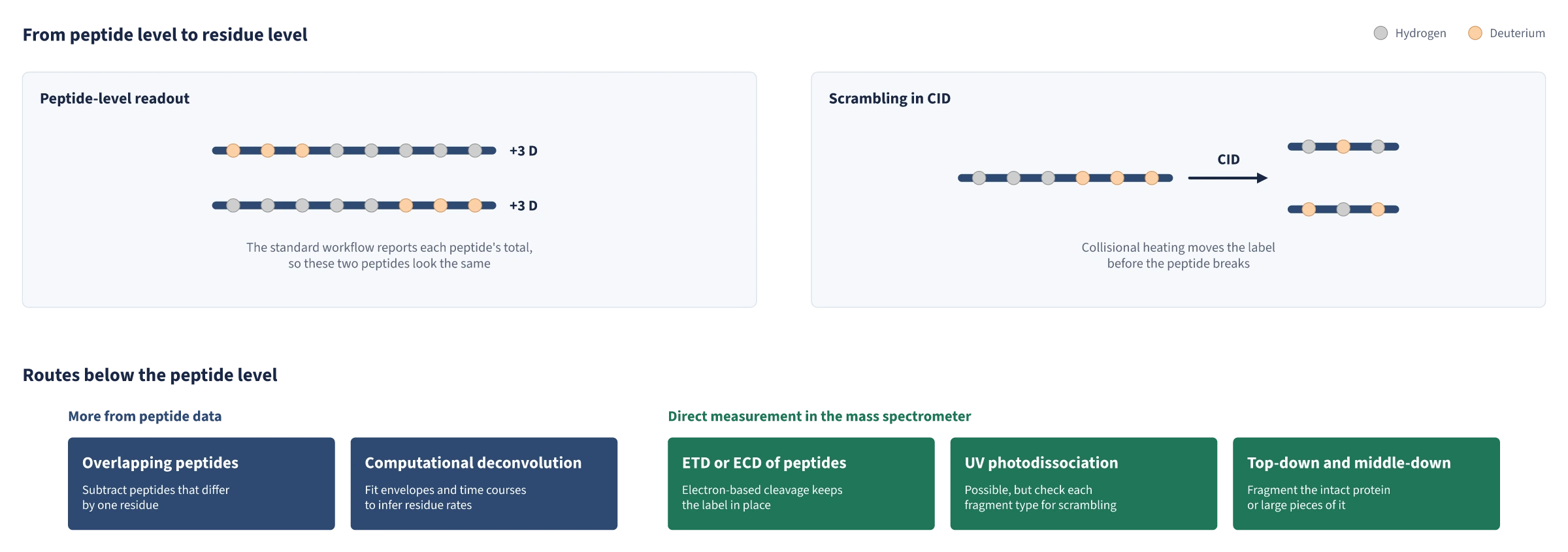
In a standard bottom-up hydrogen-deuterium exchange mass spectrometry (HDX-MS) experiment, deuterium uptake is read peptide by peptide, and spatial resolution is typically 5 or more residues, set by the length of the proteolytic peptides [1]. While many fragmentation methods are available for generating high-resolution HDX-MS data, many fail due to the scrambling of hydrogen with deuterium atoms along the peptide backbone [2]. Collision-induced dissociation (CID) is the most common fragmentation technique for breaking peptide ions into smaller fragments [1]. It uses kinetic energy to collide ions with neutral, inert gas molecules, raising the ions' internal energy and causing fragmentation [2]. Because backbone amide hydrogens are labile, heating ions during CID causes them to migrate along the peptide before fragmentation occurs [1, 3]. This scrambling erases positional information from solution labeling and can also occur due to heating in the ion source and ion optics before fragmentation [2, 3].
In drug discovery and development, many questions are asked at the level of single residues: which residues hold a ligand, whether two compounds that bind the same site bind the same way, and which residues of an epitope carry the interaction. This note reviews approaches that go beyond the peptide level, and what each one trades off.
Achieving better or single-residue resolution
There are two ways to go further. One is to extract more from peptide-level data, through more overlapping peptides or computation. The other is to break the labeled peptides, or the intact protein, inside the mass spectrometer and measure the pieces directly.
More overlapping peptides
If two peptides differ by one residue at their C-terminal end, the difference in their uptake belongs to that residue. With enough overlapping peptides, subtraction narrows deuterium down to short segments and, in some protein regions, single amides. For cytochrome c, combining proteases (pepsin with fungal protease type XIII) with different cleavage preferences produced 138 high-quality peptides, against 58 from pepsin alone, and gave exchange rates for 65 of the 100 backbone amides, consistent with an earlier published NMR study [4].
Benefits. The measurements are ordinary peptide-level data from standard instruments and workflows, with no fragmentation step in which scrambling could occur [3, 4].
Drawbacks. Resolution is uneven because it depends on where the proteases cut, and the first two residues of each peptide cannot retain deuterium, limiting subtraction from the N-terminal side [4]. Each subtraction compares peptides that lost different amounts of label during analysis, and back-exchange corrections are exact only in special cases [5]. More peptides also mean more data to curate [6].
Computational deconvolution
A peptide's isotopic envelope contains more information than its centroid mass: its shape reflects how deuterium is distributed across the peptide's amides. Fitting the envelopes of many overlapping peptides across a time course can recover exchange rates for individual residues, and in published cases, residue-level values obtained this way have agreed well with NMR measurements [7, 8]. Several tools implement this approach, including HDXmodeller, PyHDX, and ReX, and the newer ones also report how confident each residue assignment is [9, 10, 11].
Benefits. These methods work on data many laboratories already collect, require no special hardware, and cover the whole measured sequence [9, 11].
Drawbacks. The result is an inference rather than a measurement. The problem is usually underdetermined, so many residue-level models can fit the same peptide data equally well; reliable answers need high peptide redundancy and an extensive time course, which real samples do not always allow [1, 9]. Residues that never report, such as prolines, remain unresolved [9].
Electron-based fragmentation of labeled peptides
Electron capture dissociation (ECD) and electron transfer dissociation (ETD) break the peptide backbone through electron-driven reactions rather than heating. As long as ions are not heated before fragmentation, the fragments keep the solution labeling pattern [3, 12]. Applied to a digested protein, ETD gave deuterium levels for 60 individual backbone amides of β2-microglobulin that correlated closely with NMR results [13]. This approach has since been automated for differential experiments with ligands [14] and used to screen kinase inhibitors [15].
Scrambling must be checked rather than assumed absent. A scrambling probe peptide is used to tune the instrument, and the loss of ammonia during ETD indicates scrambling in the experimental peptides themselves [13, 16]. Gentle settings minimize scrambling but often do not remove it completely [2].
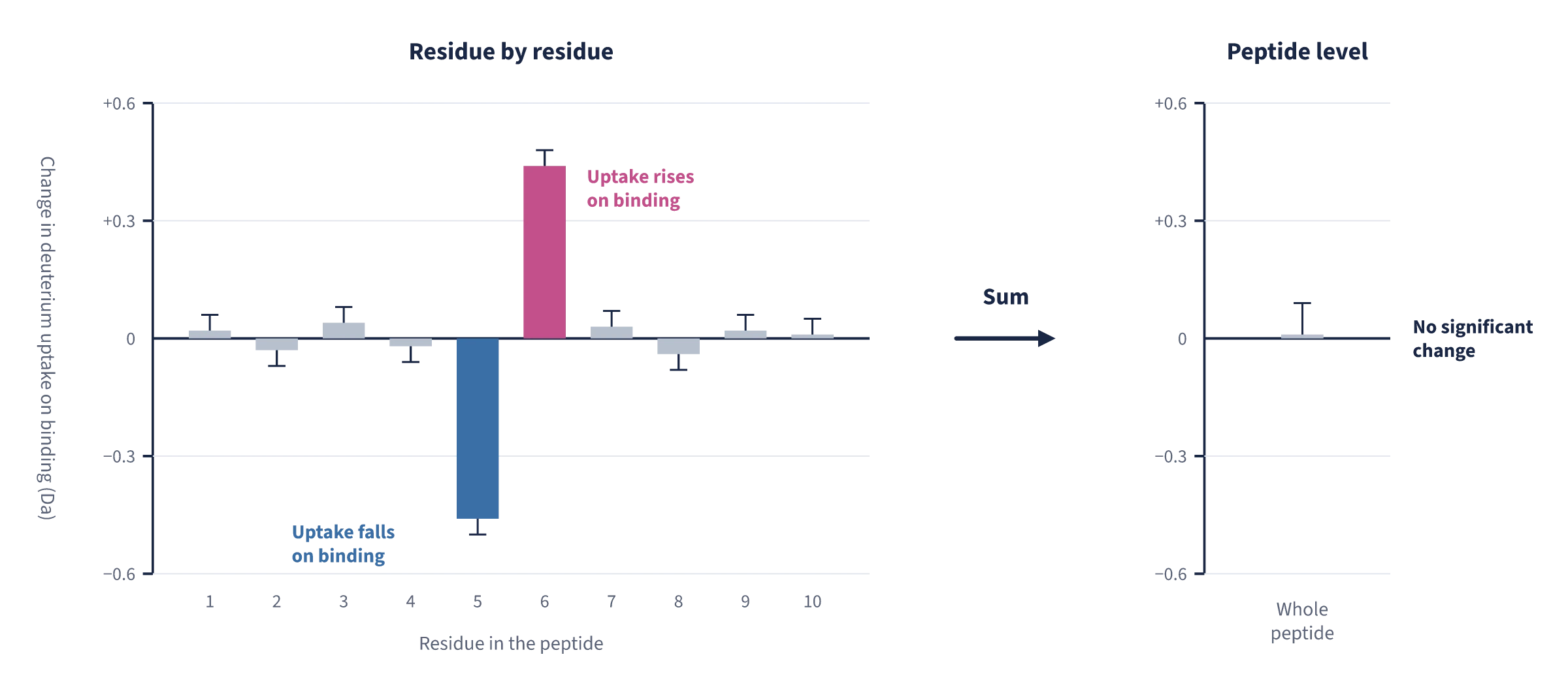
Benefits. The deuterium on each residue is measured directly rather than inferred [13], and direct measurement exposes opposite changes within a peptide that cancel in the peptide average [1, 15].
Drawbacks. The main cost is sensitivity. ECD and ETD are inefficient, losing between one and three orders of magnitude of signal relative to the parent ion, and the gentle ion transmission needed to avoid scrambling lowers it further [17, 18]. ETD works best for peptides carrying three or more charges, and not every peptide fragments well enough [15]. Software built to process ECD and ETD data is still limited [18]. Recent work on an instrument with an efficient ECD cell showed that low-scrambling conditions need not cost sensitivity, and compared ligands sharing one binding site residue by residue, although peptides still had to be chosen in advance and analyzed by hand [1, 17].
UV photodissociation
Ultraviolet photodissociation (UVPD) fragments ions with high-energy photons, causing electronic excitation that leads to bond cleavage [19].
Benefits. UVPD offers high fragmentation efficiency, and it localized deuterium without scrambling in a model peptide [20].
Drawbacks. Other work found extensive scrambling in several fragment types, so each fragment type must be validated on the instrument in use [19].
Top-down and middle-down
Instead of digesting the labeled protein, the intact protein can be fragmented by ECD or ETD. For a 17 kDa protein, this approach located protected amides with an average resolution of better than two residues while greatly reducing back-exchange [21], and top-down analysis can distinguish proteoforms that bottom-up analysis averages together [22]. For antibodies, a middle-down variant uses restricted pepsin digestion into a few large fragments; for a 150 kDa therapeutic antibody, it gave around two-residue resolution on average, single-residue resolution in many regions, and covered 96.8% of the molecule [23].
Benefits. Little or no digestion, lower back-exchange, and information on intact proteoforms [21, 22].
Drawbacks. Success falls quickly as protein size increases, and top-down HDX has mainly been applied to proteins below 30 kDa [23]. It requires very gentle source conditions, has low fragmentation efficiency, and has not been widely adopted [5].
The routes side by side
| Route | What it gives | Main benefits | Main drawbacks |
|---|---|---|---|
| More overlapping peptides | Residue values by subtraction, where cleavages allow | Standard instruments and workflows; no scrambling risk | Uneven coverage; back-exchange differences between peptides; more curation |
| Computational deconvolution | Inferred residue rates or protection factors | Uses existing data; whole measured sequence; confidence metrics in newer tools | Underdetermined; needs high redundancy and long time courses; inference, not measurement |
| ETD or ECD of peptides | Measured deuterium on individual residues | Direct; agrees with NMR | Large sensitivity loss; scrambling must be tuned out and checked; little software support |
| UV photodissociation | Potentially residue values | High fragmentation efficiency | Scrambling reported for several fragment types; less mature |
| Top-down and middle-down | Near-residue values on intact proteins or large fragments | Little digestion; low back-exchange; proteoform-specific | Falls off with protein size; gentle conditions and low efficiency; not widely adopted |
More overlapping peptides
- What it gives
- Residue values by subtraction, where cleavages allow
- Main benefits
- Standard instruments and workflows; no scrambling risk
- Main drawbacks
- Uneven coverage; back-exchange differences between peptides; more curation
Computational deconvolution
- What it gives
- Inferred residue rates or protection factors
- Main benefits
- Uses existing data; whole measured sequence; confidence metrics in newer tools
- Main drawbacks
- Underdetermined; needs high redundancy and long time courses; inference, not measurement
ETD or ECD of peptides
- What it gives
- Measured deuterium on individual residues
- Main benefits
- Direct; agrees with NMR
- Main drawbacks
- Large sensitivity loss; scrambling must be tuned out and checked; little software support
UV photodissociation
- What it gives
- Potentially residue values
- Main benefits
- High fragmentation efficiency
- Main drawbacks
- Scrambling reported for several fragment types; less mature
Top-down and middle-down
- What it gives
- Near-residue values on intact proteins or large fragments
- Main benefits
- Little digestion; low back-exchange; proteoform-specific
- Main drawbacks
- Falls off with protein size; gentle conditions and low efficiency; not widely adopted
Choosing a route
No single route resolves every residue of every protein. A sensible approach is to run a conventional peptide-level experiment first, use it to choose the regions that matter, then apply residue-level methods to those regions. Residue-level data repays the effort most where peptide averaging misleads: in binding sites where opposite changes can cancel, in comparing compounds or antibodies that share a site, and where one key amide tracks affinity better than the peptide average does.
Single-Residue HDX-MS at RaptMAb
If your project needs more than peptide-level resolution, schedule a consultation to discuss which route fits your protein and your question.
References
- 1.Anacleto J, Wolf E, Ackloo S, Arrowsmith C, LeBlanc Y, Lento C, et al. Site-specific hydrogen-deuterium exchange mass spectrometry for deep explorations of dynamic shifts in protein-drug interactions. J Am Chem Soc. 2026;148(28):29788-29794. doi:10.1021/jacs.6c03259
- 2.Mundorff CC, Hadley S, Tuttle LM, Shi Y, McAlister GC, Viner R, et al. Mapping hydrogen migration thresholds for site-specific HDX-MS. Mol Cell Proteomics. 2025;24(11):101075. doi:10.1016/j.mcpro.2025.101075
- 3.Rand KD, Adams CM, Zubarev RA, Jørgensen TJD. Electron capture dissociation proceeds with a low degree of intramolecular migration of peptide amide hydrogens. J Am Chem Soc. 2008;130(4):1341-1349. doi:10.1021/ja076448i
- 4.Hamuro Y, Zhang T. High-resolution HDX-MS of cytochrome c using pepsin/fungal protease type XIII mixed bed column. J Am Soc Mass Spectrom. 2019;30(2):227-234. doi:10.1007/s13361-018-2087-7
- 5.Konermann L, Scrosati PM. Hydrogen/Deuterium Exchange Mass Spectrometry: Fundamentals, Limitations, and Opportunities. Mol Cell Proteomics. 2024;23(11):100853. doi:10.1016/j.mcpro.2024.100853
- 6.Filandr F, Sarpe V, Raval S, Crowder DA, Khan MF, Douglas P, et al. Automating data analysis for hydrogen/deuterium exchange mass spectrometry using data-independent acquisition methodology. Nat Commun. 2024;15(1):2200. doi:10.1038/s41467-024-46610-3
- 7.Kan ZY, Walters BT, Mayne L, Englander SW. Protein hydrogen exchange at residue resolution by proteolytic fragmentation mass spectrometry analysis. Proc Natl Acad Sci U S A. 2013;110(41):16438-16443. doi:10.1073/pnas.1315532110
- 8.Stofella M, Skinner SP, Sobott F, Houwing-Duistermaat J, Paci E. High-resolution hydrogen-deuterium protection factors from sparse mass spectrometry data validated by nuclear magnetic resonance measurements. J Am Soc Mass Spectrom. 2022;33(5):813-822. doi:10.1021/jasms.2c00005
- 9.Salmas RE, Borysik AJ. HDXmodeller: an online webserver for high-resolution HDX-MS with auto-validation. Commun Biol. 2021;4(1):199. doi:10.1038/s42003-021-01709-x
- 10.Smit JH, Krishnamurthy S, Srinivasu BY, Parakra R, Karamanou S, Economou A. Probing universal protein dynamics using hydrogen-deuterium exchange mass spectrometry-derived residue-level Gibbs free energy. Anal Chem. 2021;93(38):12840-12847. doi:10.1021/acs.analchem.1c02155
- 11.Crook OM, Gittens N, Chung CW, Deane CM. Inferring residue level hydrogen deuterium exchange with ReX. Commun Chem. 2025;8(1):343. doi:10.1038/s42004-025-01719-4
- 12.Zehl M, Rand KD, Jensen ON, Jørgensen TJD. Electron transfer dissociation facilitates the measurement of deuterium incorporation into selectively labeled peptides with single residue resolution. J Am Chem Soc. 2008;130(51):17453-17459. doi:10.1021/ja805573h
- 13.Rand KD, Zehl M, Jensen ON, Jørgensen TJD. Protein hydrogen exchange measured at single-residue resolution by electron transfer dissociation mass spectrometry. Anal Chem. 2009;81(14):5577-5584. doi:10.1021/ac9008447
- 14.Landgraf RR, Chalmers MJ, Griffin PR. Automated hydrogen/deuterium exchange electron transfer dissociation high resolution mass spectrometry measured at single-amide resolution. J Am Soc Mass Spectrom. 2012;23(2):301-309. doi:10.1007/s13361-011-0298-2
- 15.Masson GR, Maslen SL, Williams RL. Analysis of phosphoinositide 3-kinase inhibitors by bottom-up electron-transfer dissociation hydrogen/deuterium exchange mass spectrometry. Biochem J. 2017;474(11):1867-1877. doi:10.1042/BCJ20170127
- 16.Rand KD, Zehl M, Jensen ON, Jørgensen TJD. Loss of ammonia during electron-transfer dissociation of deuterated peptides as an inherent gauge of gas-phase hydrogen scrambling. Anal Chem. 2010;82(23):9755-9762. doi:10.1021/ac101889b
- 17.Anacleto J, Kabir E, Blanco M, Leblanc Y, Lento C, Wilson DJ. Efficient, zero scrambling fragmentation of deuterium labeled peptides on the ZenoToF 7600 electron activated dissociation platform. J Am Soc Mass Spectrom. 2025;36(5):1175-1181. doi:10.1021/jasms.5c00041
- 18.Anacleto J, Ackloo S, Arrowsmith C, Wolf E, Leblanc Y, Lento C, et al. Site-specific hydrogen deuterium exchange difference mass spectrometry measurements for ligand binding. J Am Soc Mass Spectrom. 2025;36(10):2315-2318. doi:10.1021/jasms.5c00229
- 19.Modzel M, Wollenberg DTW, Trelle MB, Larsen MR, Jørgensen TJD. Ultraviolet photodissociation of protonated peptides and proteins can proceed with H/D scrambling. Anal Chem. 2021;93(2):691-696. doi:10.1021/acs.analchem.0c02957
- 20.Mistarz UH, Bellina B, Jensen PF, Brown JM, Barran PE, Rand KD. UV photodissociation mass spectrometry accurately localize sites of backbone deuteration in peptides. Anal Chem. 2018;90(2):1077-1080. doi:10.1021/acs.analchem.7b04683
- 21.Pan J, Han J, Borchers CH, Konermann L. Hydrogen/deuterium exchange mass spectrometry with top-down electron capture dissociation for characterizing structural transitions of a 17 kDa protein. J Am Chem Soc. 2009;131(35):12801-12808. doi:10.1021/ja904379w
- 22.Langford JB, Ahmed E, Fang M, Cupp-Sutton K, Smith K, Wu S. Strategies for top-down hydrogen deuterium exchange-mass spectrometry: a mini review and perspective. J Mass Spectrom. 2024;59(11):e5097. doi:10.1002/jms.5097
- 23.Pan J, Zhang S, Chou A, Borchers CH. Higher-order structural interrogation of antibodies using middle-down hydrogen/deuterium exchange mass spectrometry. Chem Sci. 2016;7(2):1480-1486. doi:10.1039/c5sc03420e